Cleaved Caspase 3 Immunofluorescence
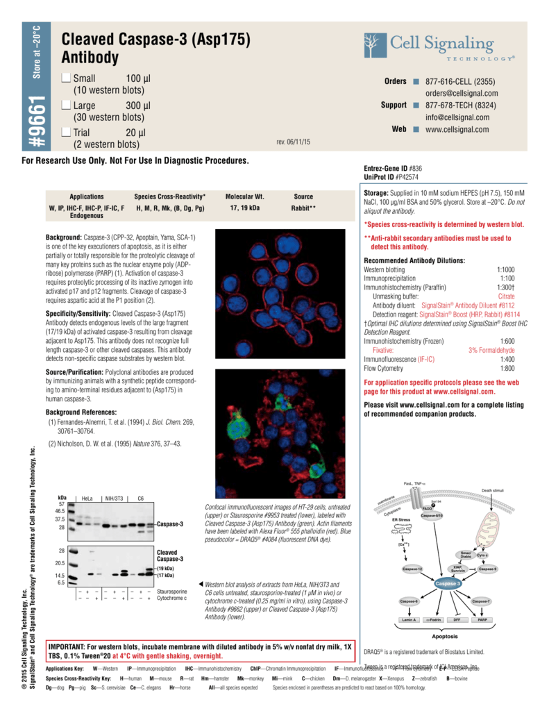
Blue pseudocolor = DRAQ5 ® #4084 (fluorescent DNA dye).
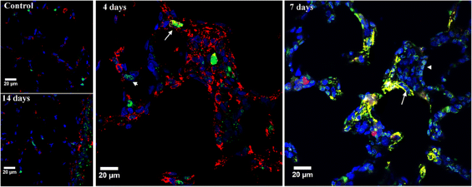
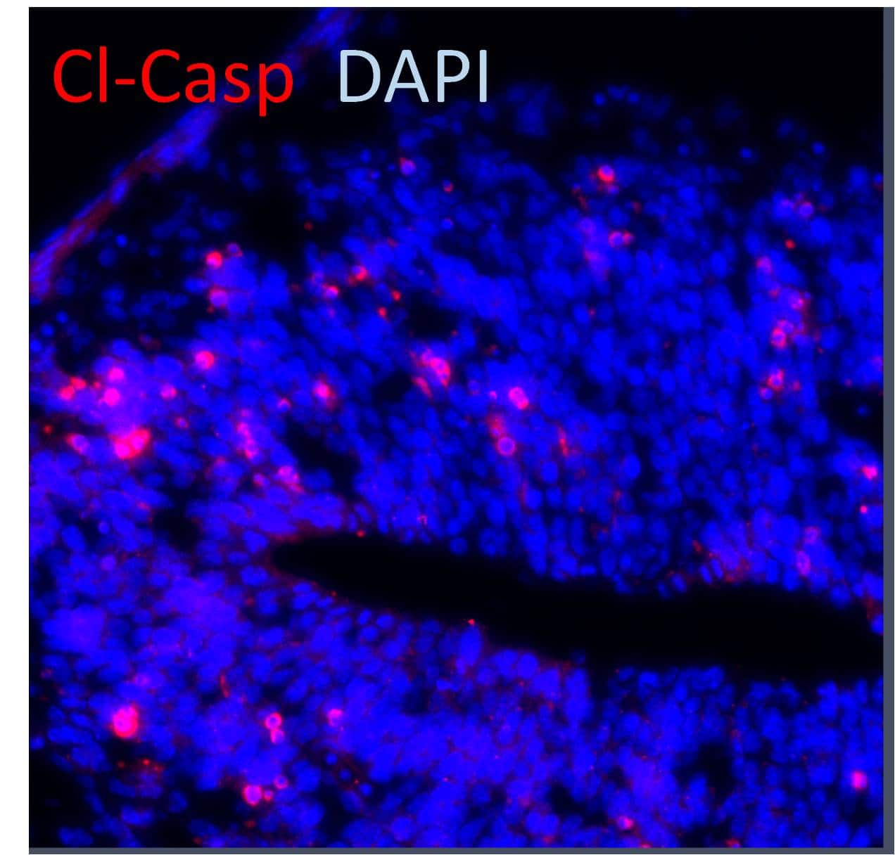
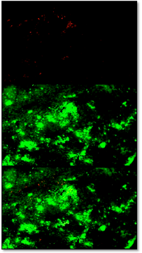
Cleaved caspase 3 immunofluorescence. The immunofluorescence data demonstrate that there is limited association of cleaved-caspase-3-positive puncta with EBA-immunopositive cells, despite the presence of an abundance of cleaved-caspase-3-positive puncta and diffuse immunoreactivity both extracellularly and intracellularly (i.e., co-localized with DAPI) in the surrounding areas (see excerpt (2) in Figure 2). AA6, monoclonal ChIP, IHC, IP, WB pig, rat, human, mouse, rat, rabbit, chicken, rabbit, pig, guinea. Flow Cytometry (FCM) General Protocol.
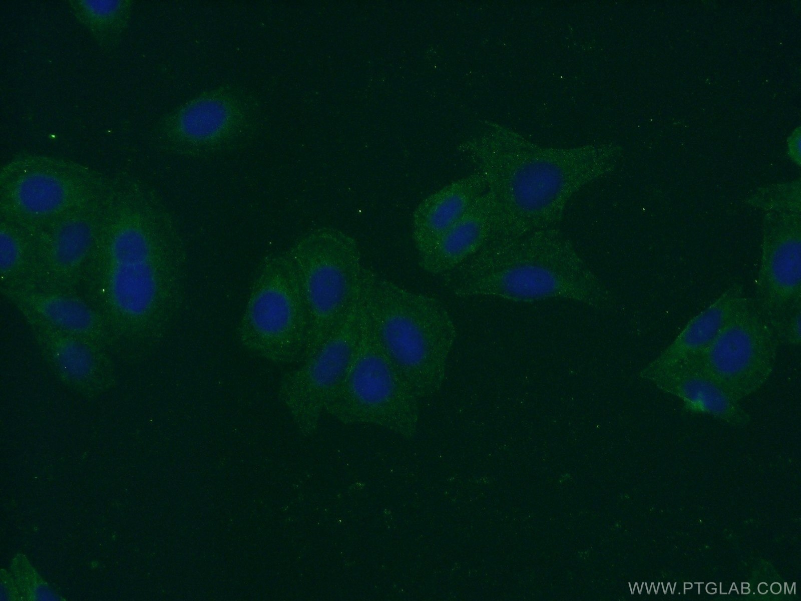
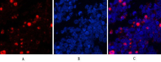
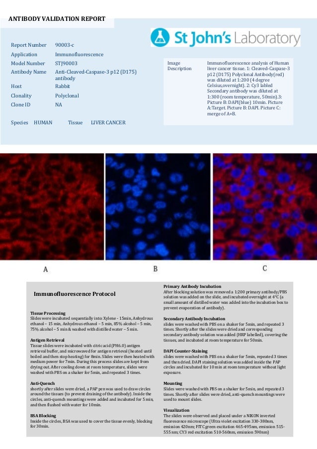
Cleaved Caspase-3 (Asp175) Antibody, 9661S - Get the Best Quote/Price and read Reviews, Features and Research Applications. 2, Cy3 labled Secondary antibody was diluted at 1:300(room temperature, 50min).3, Picture B:. The p43 is then cleaved to yield p26 and the release of the active site containing p18.
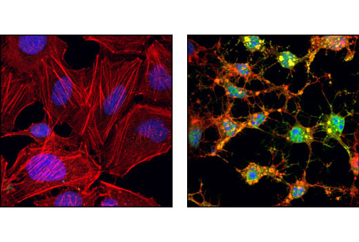
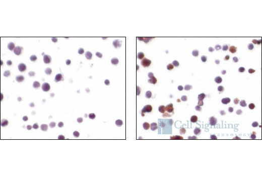
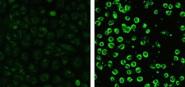
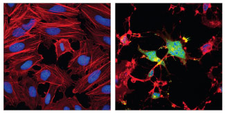
Staining of the treated cells with anti-ki-67 and anti-cleaved caspase-3 (Abcam, Cambridge, MA) was performed overnight at 4 °C in PBS containing 1% BSA and 0.1% tween. Confocal immunofluorescent images of HT-29 cells, untreated (left) or Staurosporine #9953 treated (right) labeled with Cleaved Caspase-3 (Asp175) (5A1E) Rabbit mAb (green). The cleavage of caspase-8 to generate a 43 and a 12 kDa fragment which is further processed to 10 kDa.
(Strsp) from <i>Streptomyces</i> sp. 8/13/ Rabbit anti-Caspase-3 cleaved (Asp175) Antibody for Immunofluorescence José Vieira This antibody works really well to detect cells in apoptosis during development. For 6 hrs served as a positive control.
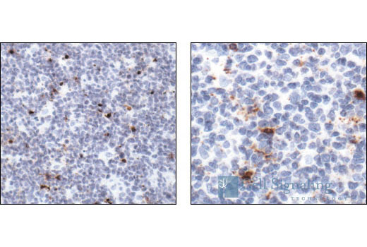
Sections of germinal centers of normal lymph node seen at low (A,B) and high (C-F) magnification. *P < 0.05, **P < 0. Cleaved Caspase-3 (Asp175) (5A1) Rabbit mAb detects endogenous levels of the large fragment (17/19 kDa) of activated caspase-3 resulting from cleavage adjacent to Asp175.
This antibody detects non-specific caspase substrates by western blot. The lane on the left is treated with the synthesized peptide. Activation of caspase-8 involves a two-step proteolysis:.
Data are expressed as the mean ± SD (n = 5), and were analyzed by one-way analysis of variance. Immunocytochemistry / Immunofluorescence (3) Western Blot (3) ELISA (2) Enzyme Activity. Caspases 6, 7 and 9), as well as relevant targets in the cells (e.g.
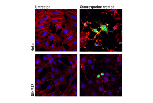
2, Cy3 labled Secondary antibody was diluted at 1:300(room temperature, 50min).3, Picture B:. Confocal immunofluorescent analysis of HeLa cells, untreated (left) or treated with Staurosporine #9953 (1 μM, 4 hr;. I 've been having some issues with detecting cleaved caspase 3, 7 and 9 in my w.blots.
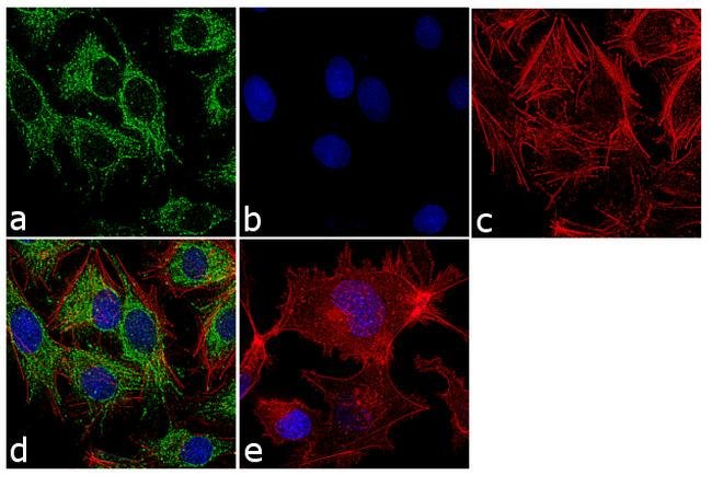
The issue with Caspase staining also is that. Immunocytochemistry/ Immunofluorescence - Anti-Caspase-3 antibody (ab) This image is courtesy of Roberto Giambruno, Marilena Ciciarello and Patrizia Lavia HeLa cells were fixed for 10 minutes at room temperature in 3.7% PFA and permeabilised in 0.1% Triton X-100/PBS then incubated with ab (5µg/ml) for 1 hour at room temperature. The cells were fixed with 4% paraformaldehyde for 10 minutes, permeabilized with 0.1% Triton™ X-100 for 15 minutes, and blocked with 1% BSA for 1 hour at room temperature.
Actin filaments have been labeled with Alexa Fluor® 555 phalloidin #53 (red). Search results for cleaved caspase 3 at Sigma-Aldrich. Immunohistochemistry to active caspase-3, recently recommended for apoptosis detection, is inappropriate to detect apoptosis involving caspase-7.
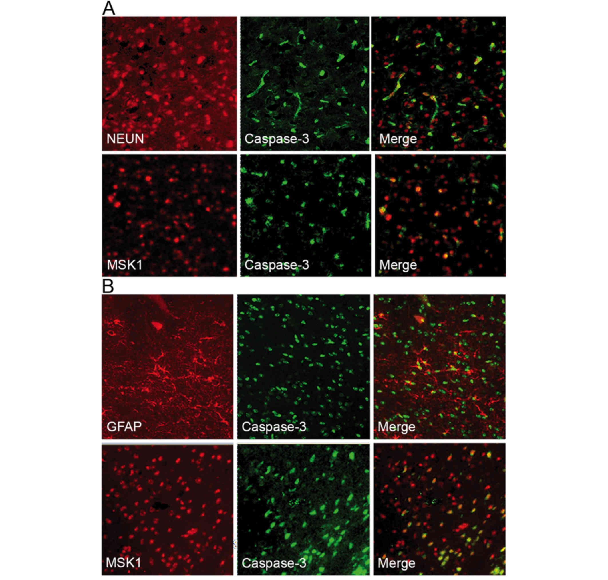
Anti-Caspase 3 Antibody, active (cleaved) form detects level of Caspase 3 and has been published and validated for use in Immunofluorescence (IF), Immunohistochemistry (IHC), and Western Blot (WB). The cleaved caspase-3 also collocated with microglia and astrocytes indicating its participation in glial activation. 219.0 - 14,000 pg/mL.
Several research groups have reported that LMW RSNOs impede caspase 3 activity via reactions of trans-S-nitrosation 9, 10, 12, 13, 31, 35, 36.Detailed mechanistic studies by Zech et al. Anti-Caspase 3 Antibody, active (cleaved) form detects level of Caspase 3 and has been published and validated for use in Immunofluorescence (IF), Immunohistochemistry (IHC), and Western Blot (WB). Immunofluorescence and fluorescently tagged Atg8 can be used to visualize the protein localization and autophagy activity of a cell by microscopy.
Lay the slides flat in a humidified chamber, protected from light, and incubate for 1-2 hr at room temperature. Cleaved-Caspase-9 (D353) Antibody was diluted at 1:0(4°C,ON). Colocalization obtained from merged immunofluorescence images indicated that active caspase-7 was not expressed in all caspase-3–expressing cells , suggesting that Foscan-PDT–induced apoptosis was mainly processed through the activation of caspase-3 in HT29 tumors.
Figure 6 Cleaved caspase 3, myogenin and vimentin in the gastrocnemius muscles 12 weeks after ADSC transplantation (western blot assay). Caspase 3/p17/p19 antibody Mouse Monoclonal from Proteintech validated in Western Blot (WB), Immunohistochemistry (IHC), Immunofluorescence (IF), Enzyme-linked Immunosorbent Assay (ELISA) applications. Specific anti-(Asp175) cleaved caspase-3 primary antibodies and Alexa Fluor 594 secondary antibodies were used to visualize activated caspase-3 (first column), and nuclei were.
Have established that caspase 3 can undergo poly-S-nitrosation, whereby all S-nitroso functions in the p12 subunit are cleaved with the release of NO and partial formation of protein-mixed disulfides with GSH. Because PKCδ possesses a caspase-3–specific cleavage site (DMQD 329 /N) at the hinge region that is critical to caspase-3 and caspase-7–mediated cleavage and activation , we transduced the DN_PKCδ (D329A) mutant into the HT-29 and HCT116 cells (Fig. The cells were harvested after treatment and were stained with JC-1.
Monoclonal antibody is produced by immunizing animals with a synthetic peptide corresponding to amino-terminal residues adjacent to Asp175 of human caspase-3. Caspase-3 is a caspase protein that interacts with caspase-8 and caspase-9. Host Goat Type Primary Modification Cleaved.
Cleaved-Caspase 3 antibody Rabbit Polyclonal from Proteintech validated in Western Blot (WB), Immunoprecipitation (IP), Immunohistochemistry (IHC), Immunofluorescence (IF), Flow Cytometry (FC), Enzyme-linked Immunosorbent Assay (ELISA) applications. Caspase-3, Apopain, Cysteine protease CPP32, Protein Yama, SREBP cleavage activity 1, CASP3_HUMAN. Caspase-3 cleaves multiple substrates including PARP, proIL-16, PKC-gamma and -delta, proCaspases-6, -7, and -9, and beta-Catenin.
During apoptosis, however, proCaspase-3 is activated by cleavage into p and p12 subunits, and the p subunit is trimmed to yield a p17 subunit. Immunofluorescence IF-Fr Immunofluorescence-frozen IF-P Immunofluorescence-paraffin IF-WM Immunofluorescence-wholemount IHC Immunohistochemistry. Right), using Cleaved Caspase-3 (Asp175) (D3E9) Rabbit mAb (Alexa Fluor ® 647 Conjugate) (blue pseudocolor).
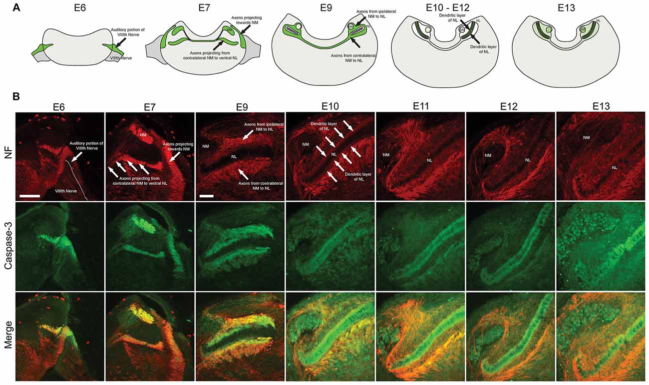
Cleaved-Caspase-9 (D353) Antibody(red) was diluted at 1:0(4°C,ON). D Quantitation of Cleaved caspase-3 and Cytochrome C levels in U251 cells treated. For example, caspase activity can be visualized through the loss of nuclear Lamins and an increase in processed (cleaved) Caspase-3 in salivary glands (Fig.
1,Cleaved-Caspase-3 p12 (D175) Polyclonal Antibody(red) was diluted at 1:0(4°C,overnight). Actin filaments were labeled with Alexa Fluor ® 4 Phalloidin #78 (green). Immunofluorescence analysis of Caspase 9 (Cleaved Asp315) was performed using 70% confluent log phase HeLa cells treated with 1µM of Staurosporine for 3 hours.
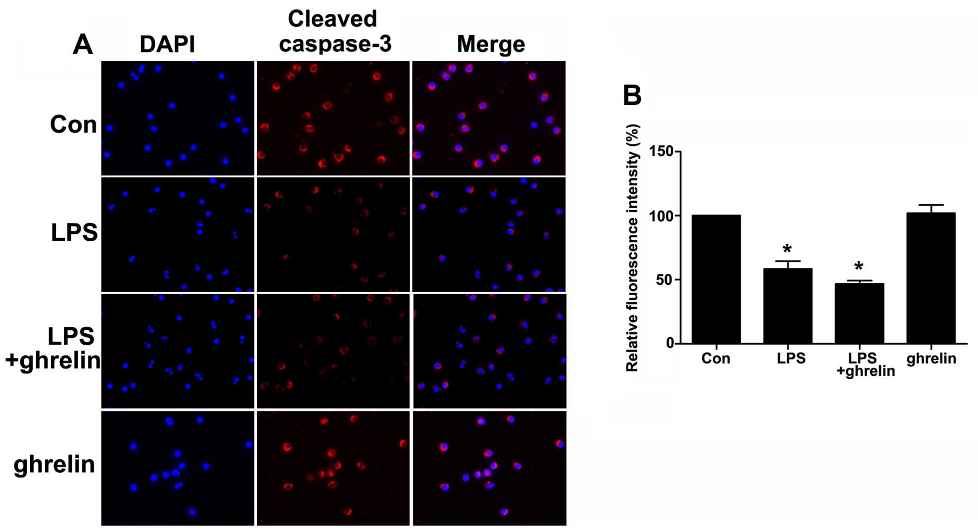
This antibody does not recognize full length caspase-3 or other cleaved caspases. Immunofluorescence analysis of cleaved caspase-3 levels in treated microglial cells. Variations in levels of Caspase 3 have been reported in cells of short-lived nature and those with a longer life cycle.
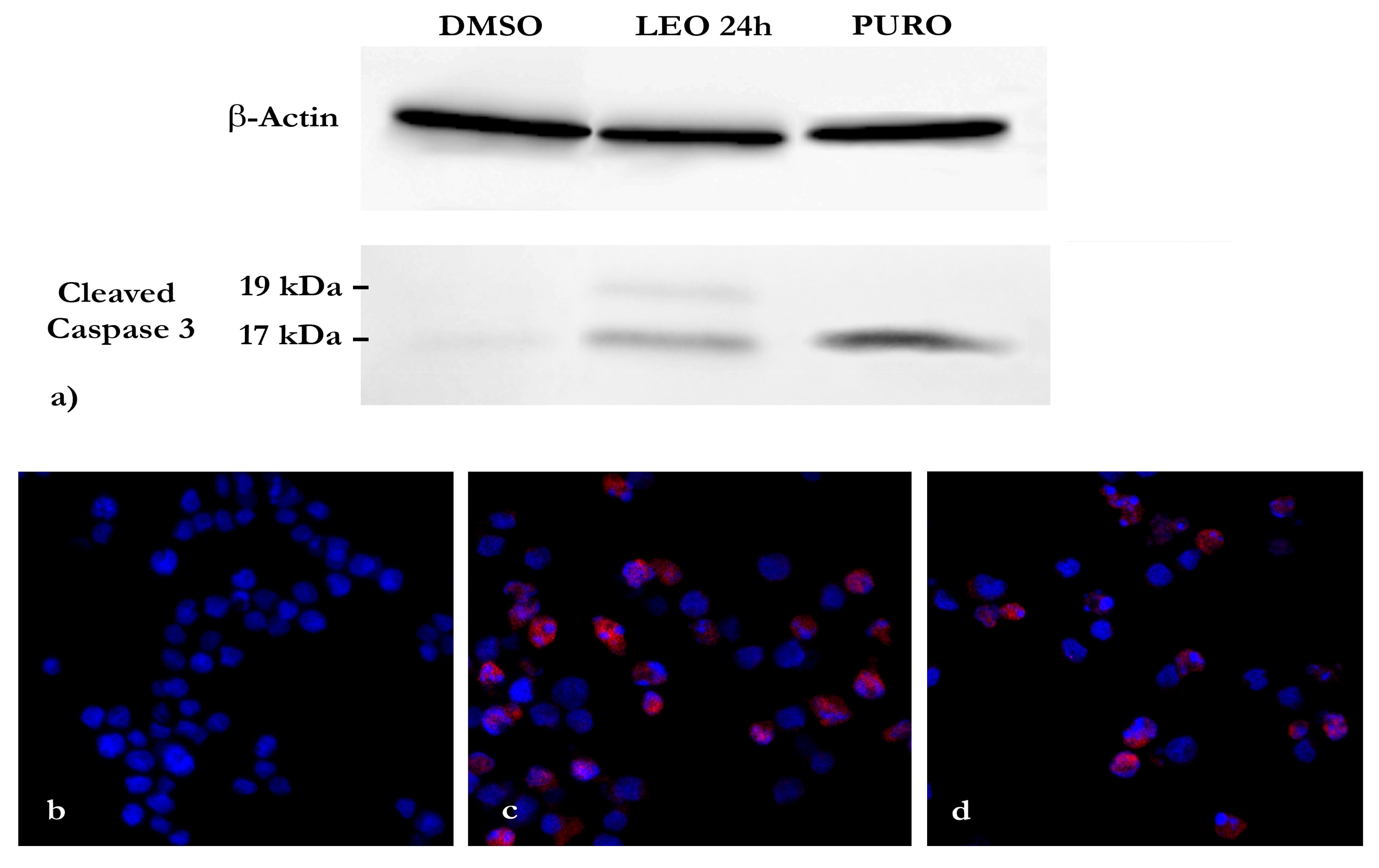
This antibody reacts with human, mouse, rat samples. C Western blot analysis for the expressions of Caspase-3, Cleaved caspase-3, and Cytochrome C in U251 cells treated with control (DMSO) or 3-MA (5 mM) with or without bortezomib (10 nM) for 24 h. Cleaved Caspase-3 (Asp175) IHC Detection Kit rev.
Immunofluorescence analysis of Caspase-3 was done on 70% confluent log phase HeLa cells treated with 5 uM of Staurosporine for 16 hours. Apoptosis evaluation induced in …. Cleaved Caspase-3 (Asp175) Antibody detects endogenous levels of the large fragment (17/19 kDa) of activated caspase-3 resulting from cleavage adjacent to Asp175.
Caspase 3 is cleaved at Asp28/Ser29 and Asp175/Ser176. Immunofluorescence(IC) Immunohistochemistry(Polymer) Western Blotting. (sc-) cleaved caspase-3 p11 Antibody (h176) Antibody info;.
Products by Category. Skip to main content. Research CategoryApoptosis & CancerMetabolism.
One of these is that present on activated caspase 3, a ubiquitously distributed caspase that is a main effector caspase of the apoptotic cascade within cells. Normally, it is an inactive, exclusively cytosolic homodimer. Target protein levels are presented as the ratio of the optical density to that of GAPDH.
Supplier Santa Cruz Biotechnology. Western blot analysis of extracts from COLO cells, using Caspase 3 (p17,Cleaved-Asp175) Antibody. Wash three times in PBS/0.1% Tween for 5 min, protected from light.
Immunofluorescence analysis of Human-liver-cancer tissue. For cleaved caspase-3 immunofluorescence we used a rabbit polyclonal antibody (Cell Signaling Technology #9661, Danvers, MA, USA) and a rabbit monoclonal antibody (R&D Systems #AF5, Minneapolis, MN, USA) that were generated using distinct synthetic peptides corresponding to human cleaved caspase-3 diluted 1:0 in blocking solution. This association is mostly reflected by adjacent direct localization of cleaved-caspase-3-positive puncta and/or DAPI.
Research Sub CategoryCaspasesEnzymes & Biochemistry. This study demonstrates the utility of using a recently commercially available antibody to cleaved caspase 3 in archival paraffin sections, suggesting that this may be a highly specific. The cells were labeled with Caspase-3 (9H19L2) Recombinant Rabbit Monoclonal Antibody ( Product # 7001) at 1 µg/mL in 0.1% BSA and incubated for 3 hours at room temperature and then.
This antibody detects non-specific caspase substrates by western blot. This antibody does not recognize full length caspase-3 or other cleaved caspases. The cells were fixed with 4% paraformaldehyde for 10 minutes, permeabilized with 0.1% Triton™ X-100 for 10 minutes, and blocked with 5% BSA for 1 hour at room temperature.
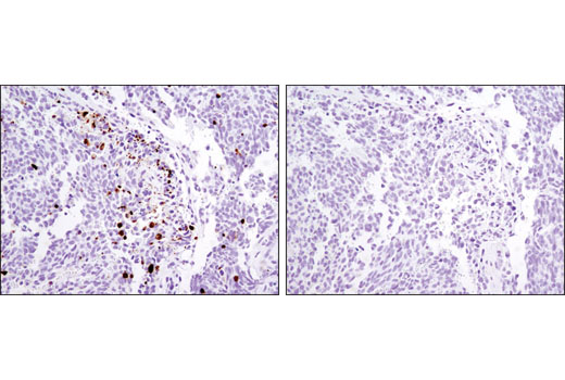
SignalStain® Cleaved Caspase-3 (Asp175) IHC Detection Kit is a “ready to use” system designed to detect the activation of caspase-3 in human tissue and cell preps by immunohistochemistry. The active Caspase 3 proteolytically cleaves and activates other caspases (e.g. This antibody reacts with Human, mouse, rat samples.
Negative control was defined by secondary antibody only. Unique orthologs are also present in birds, lizards, lissamphibians, and teleosts. Cleaved Caspase-3 Immunofluorescence Antibodies We study PyMT driven breast tumor.
1,Cleaved-Caspase-3 p17 (D175) Polyclonal Antibody(red) was diluted at 1:0(4°C,overnight). A8K5M2, D3DP53, P, Q96AN1, Q96KP2. Caspase-3 (CPP-32, Apoptain, Yama, SCA-1) is one of the key executioners of apoptosis, as it is either partially or totally responsible for the proteolytic cleavage of many key proteins such as the nuclear enzyme poly (ADPribose) polymerase (PARP) (1).
Actin filaments have been labeled with Alexa Fluor® 555 phalloidin (red). Immunofluorescence analysis of human-stomach-cancer tissue. CASP3 orthologs have been identified in numerous mammals for which complete genome data are available.
Cleaved caspase-9 further processes other caspase members, including caspase-3 and caspase-7, to initiate a caspase cascade, which leads to apoptosis (7-10). Confocal immunofluorescent analysis of NIH/3T3 cells, staurosporine-treated (left) or untreated (right), using Cleaved Caspase-9 (Asp353) Antibody (Mouse Specific) (green). This antibody does not recognize full length caspase-3 or other cleaved caspases.
01//12 n 1 Kit (150 slides) Description:. To elaborate, I am working with A375 (human melanoma), SQB (human squamous h&n carcinoma), HT1080 (human. Immunofluorescence experiments further showed cellular co-localization of cleaved-caspase-3 with GFAP and CD68 and its adjacent localization with EBA, suggesting involvement of apoptosis and neuroinflammation in mechanisms of delayed BBB and microvascular damage that could contribute to white matter changes.
It is encoded by the CASP3 gene. We chose this a-CC3 antibody due to its reliability. Yet, we also want to understand epithelial cell biology in normal mammary gland development.
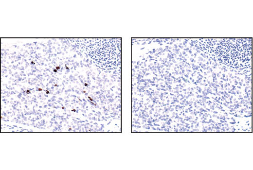
PIWI-Interacting RNA- Is Regulated by S1P Receptor Signaling Pathway to Keep Myeloma Cell Survival.In Front Oncol on Apr 15 by Ma H, Wang H, et alPMID:. Hematoxylin and eosin-stained sections (A,C,E) compared with sections immunostained with antibodies to cleaved caspase 3 (B,D,F). Cleaved Caspase-3 (Asp175) Antibody detects endogenous levels of the large fragment (17/19 kDa) of activated caspase-3 resulting from cleavage adjacent to Asp175.
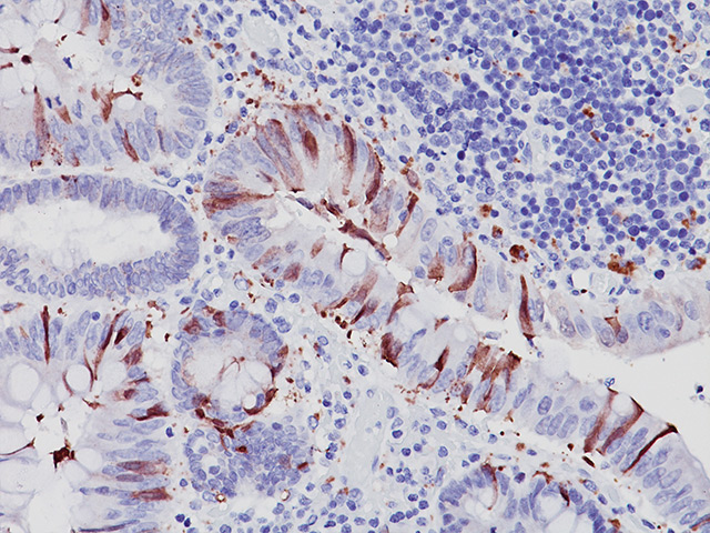
Another cleavage occurs at Asp330 producing a p37 subunit that can serve to amplify the apoptotic response (3-6). Immunohistochemical analysis of paraffin-embedded Human-liver-cancer tissue. Figure 3 Localization of cleaved caspase 3 in deparaffinized sections of formalin-fixed tissues.
Likewise, MDA-MB231 monolayer cells could be more susceptible to caspase-3. - Find MSDS or SDS, a COA, data sheets and more information. Red = Propidium Iodide (PI)/RNase Staining Solution #4087.
Human/Mouse Cleaved Caspase-3 (Asp175) DuoSet IC ELISA. View R&D Systems research products for Caspase-3. Blue pseudocolor = DRAQ5® #4084 (fluorescent DNA dye).
Our results suggest that caspase-3 and GSK-3β pY216 activation might participate in the DA cell death and that the active caspase-3 might also participate in the neuroinflammation caused by the striatal 6-OHDA injection. Immunofluorescence analysis of Human-lung-cancer tissue. ( B ) Ki-67 and cleaved caspase-3 immunofluorescence of BGC-3 cells exposed to equivalent doses of Ptx/Tet, P/T-NPs or P/T-NPs-Gelatin for 48 h.
Drain the liquid, mount the slides in a permanent or aqueous mounting medium and observe with a fluorescence microscope.

Immunofluorescent Staining Of Cleaved Caspase 3 A And Map 2 B Download Scientific Diagram

Sensory Neurons With Activated Caspase 3 Survive Long Term Experimental Diabetes Diabetes
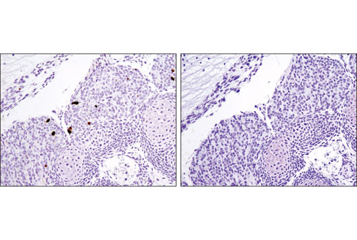
Signalstain Apoptosis Cleaved Caspase 3 Ihc Detection Kit
Cleaved Caspase 3 Immunofluorescence のギャラリー

Validated Anti Cleaved Caspase 9 P35 D315 Antibody Antibodyplus Antibody Trial And Validation
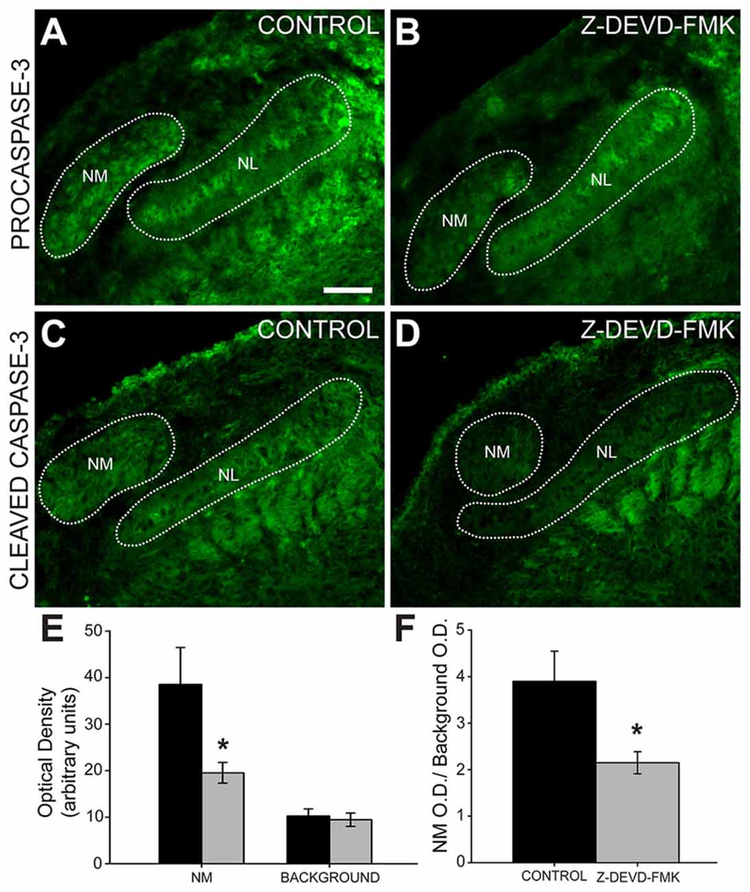
Frontiers Axonal Cleaved Caspase 3 Regulates Axon Targeting And Morphogenesis In The Developing Auditory Brainstem Frontiers In Neural Circuits

Detection Of Active Caspase 3 In Mouse Models Of Stroke And Alzheimer S Disease With A Novel Dual Positron Emission Tomography Fluorescent Tracer 68ga Ga Tc3 Ogdota
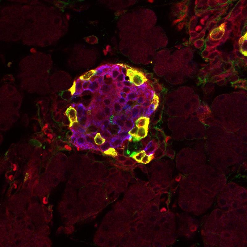
Cleaved Caspase 3 Asp175 Antibody Cell Signaling Technology

Numa And Nuclear Lamins Behave Differently In Fas Mediated Apoptosis Journal Of Cell Science

Cleaved Caspase 3 Asp175 Antibody 9661

Jci Hiv Protease Inhibitors Provide Neuroprotection Through Inhibition Of Mitochondrial Apoptosis In Mice

Anti Cleaved Caspase 3 Antibody Ab2302 Abcam

Xmlinkhub
Cleaved Caspase 3 Antibody 1 Ap Proteintech
Cleaved Caspase 3 Asp175 D3e9 Rabbit Mab 9579
Pro Caspase 3 Antibody Ma1

Cleaved Caspase 3 Asp175 Antibody 9661

Anti Casp3 Antibody Rabbit Cleaved Caspase 3 Polyclonal Antibody Np 2
Anti Caspase 3 Active Antibody Activated Caspase 3 Antibody Sigma Aldrich

Acute Lung Injury By Gastric Fluid Instillation Activation Of Myofibroblast Apoptosis During Injury Resolution Respiratory Research Full Text
Plos One Pathological Alterations In Liver Injury Following Congestive Heart Failure Induced By Volume Overload In Rats

Caspase 1 And 3 Are Sequentially Activated In Motor Neuron Death In Cu Zn Superoxide Dismutase Mediated Familial Amyotrophic Lateral Sclerosis Pnas

Anti Cleaved Caspase 3 Antibody Ab2302 Abcam
Effects Of Ghrelin On The Apoptosis Of Human Neutrophils In Vitro

Recombinant Anti Cleaved Caspase 3 Antibody E 77 Ko Tested Ab342 Abcam

Immunofluorescence An Overview Sciencedirect Topics

Cleaved Caspase 3 Biocare Medical Bioz Ratings For Life Science Research
Cleaved Caspase 3 Asp175 5a1e Rabbit Mab 9664

Enhanced Caspase Activity Contributes To Aortic Wall Remodeling And Early Aneurysm Development In A Murine Model Of Marfan Syndrome Arteriosclerosis Thrombosis And Vascular Biology

Bortezomib Induced Expression Of Chop And Cleaved Caspase 3 In Both Download Scientific Diagram

Figure S3 Multiple Staining Using The Activated Caspase 3 Antibody And Download Scientific Diagram
Cleaved Caspase 3 Asp175 Antibody Alexa Fluor 4 Conjugate

Validated Anti Cleaved Caspase 3 P12 D175 Antibody Antibodyplus Antibody Trial And Validation

Caspase 3 Antibody 85 3 Knockout Validated Products In Validated Antibody Database 101 Cited In The Literature 398 Total From 35 Suppliers

Anti Caspase 3 Antibody Active Cleaved Form Clone Chemicon From Rabbit Sigma Aldrich
Human Mouse Cleaved Caspase 3 Asp175 Antibody Mab5 R D Systems

Anti Caspase 3 Antibody Products Biocompare
Caspase 3 Polyclonal Antibody Bioss
Q Tbn 3aand9gcs2fe2gkdunlakufiorpmkspa Ijps Xeri1vavadfqfs7cdpoa Usqp Cau
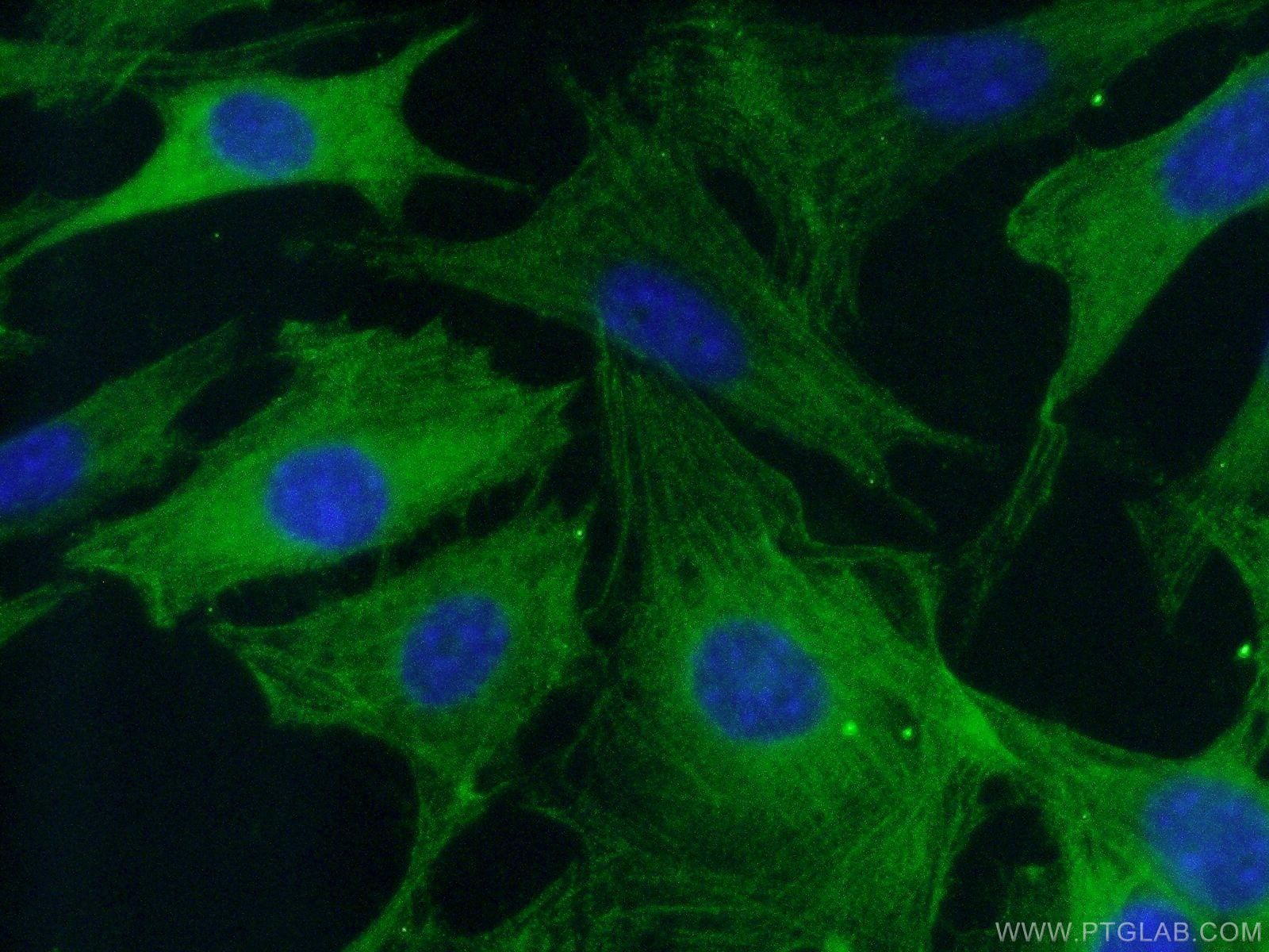
Kd Ko Validated Caspase 3 Antibody 1 Ap Proteintech
Caspase 3 Active Or Casp3 Antibody
Cleaved Caspase 3 Asp175 Antibody
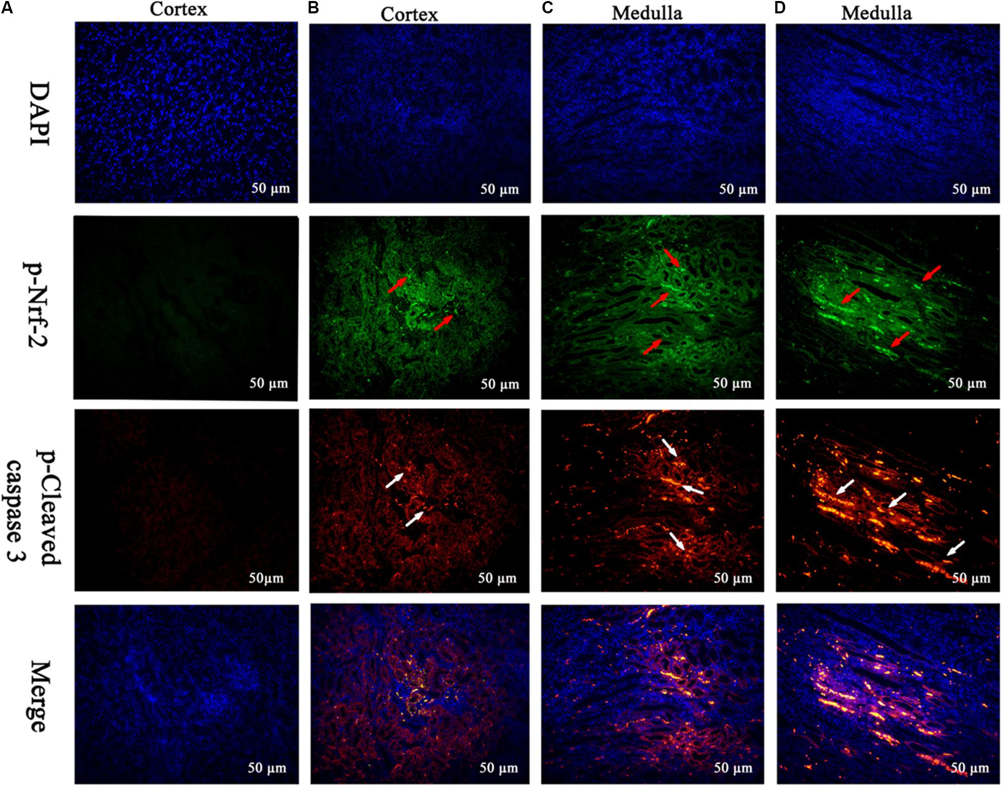
Frontiers Mequindox Induced Kidney Toxicity Is Associated With Oxidative Stress And Apoptosis In The Mouse Pharmacology

Recombinant Anti Caspase 3 Antibody E87 Ko Tested Ab Abcam

Anti Cleaved Caspase 3 Antibody Ab2302 Abcam

Immunofluorescence Assay Of Trpm7 And Cleaved Caspase 3 Expression In Download Scientific Diagram
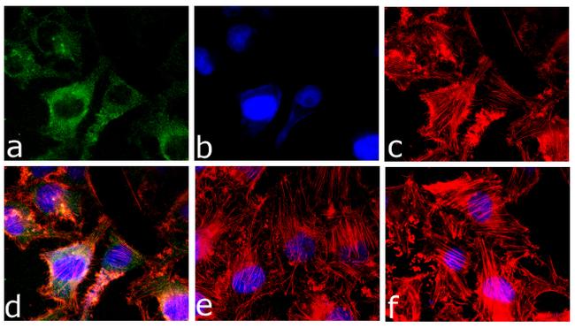
Caspase 3 Antibody 7001

Ihc World Image Gallery User Galleries
2
Human Mouse Active Caspase 3 Antibody Af5 R D Systems
Cleaved Caspase 3 Asp175 5a1e Rabbit Mab 9664

Immunoexpression Of Cleaved Caspase 3 Shows Lower Apoptotic Area Indices In Lip Carcinomas Than In Intraoral Cancer
-Immunohistochemistry-Paraffin-NB100-56113-img0004.jpg)
Caspase 3 Antibody Active Cleaved Nb100 Novus Biologicals

Fas Fasl And Cleaved Caspases 8 And 3 In Glioblastomas A Tissue Microarray Based Study Sciencedirect
---(Pro-and-Active)-Immunohistochemistry-Paraffin-NB100-56708-img0042.jpg)
Caspase 3 Antibody 31a1067 Pro And Active Nb100 Novus Biologicals
Caspase 3 Antibody 7001
Www Cell Com Molecular Cell Pdf S1097 2765 18 0 Pdf
Molecules Free Full Text Apoptotic Effects On Hl60 Human Leukaemia Cells Induced By Lavandin Essential Oil Treatment Html
Beta Static Fishersci Com Content Dam Fishersci En Us Documents Programs Scientific Technical Documents Application Notes Caspase 3 Activiation Indicator Apoptosis Application Notes Pdf

View Image
Caspase 3 Antibody Bicoare Medical
Frontiers Axonal Cleaved Caspase 3 Regulates Axon Targeting And Morphogenesis In The Developing Auditory Brainstem Frontiers In Neural Circuits
Cleaved Caspase 3 Asp175 Antibody

Jci Identification Of Thrombospondin 1 Tsp 1 As A Novel Mediator Of Cell Injury In Kidney Ischemia
Caspase 3 Signaling Cell Signaling Technology
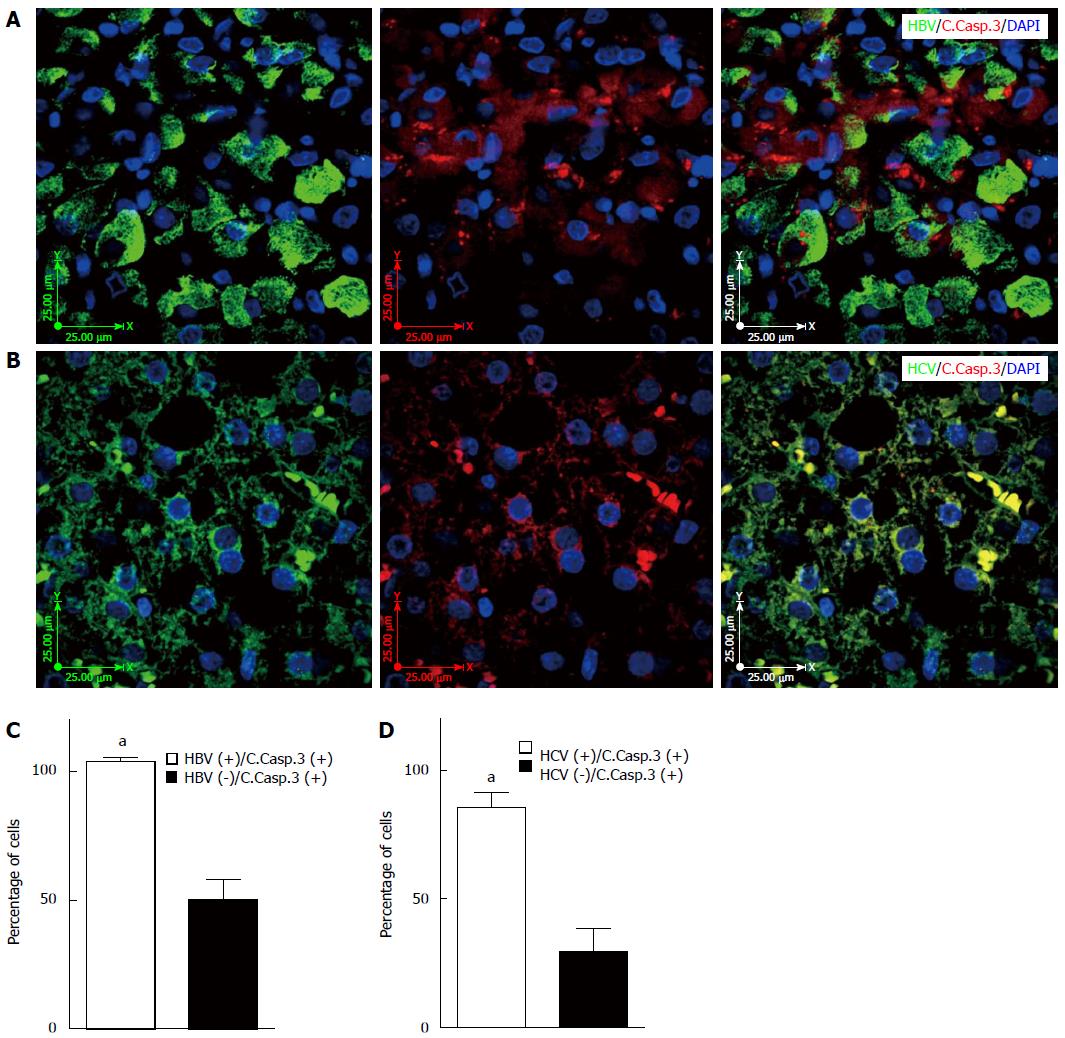
Hepatitis B And C Virus Induced Hepatitis Apoptosis Autophagy And Unfolded Protein Response

Detection Of Apoptotic Keratinocytes In A Case Of Bullous Pemphigoid Developed After Graft Versus Host Disease Html Acta Dermato Venereologica

Cleaved Caspase 3 Antibody For Immunofluorescence Biocompare Antibody Review
Jvi Asm Org Content 81 6 2614 Full Pdf
Http Histochemistry Eu Pdf Taatjes08 Pdf

In Vivo Evidence That Caspase 3 Is Required For Fas Mediated Apoptosis Of Hepatocytes The Journal Of Immunology

Anti Casp3 Antibody Rabbit Caspase 3 Polyclonal Antibody Np 2

The Detection Of Cleaved Caspase 3 In Adult Peripheral Blood Download Scientific Diagram

Caspase 3 Antibody Unconjugated Cleaved Asp175 Mab5 Novus Biologicals

Human Mouse Cleaved Caspase 3 Asp175 Antibody Mab5 R D Systems

Caspase 3 Modulates Regenerative Response After Stroke Fan 14 Stem Cells Wiley Online Library

Functional Role Of Caspase 1 And Caspase 3 In An Als Transgenic Mouse Model Science
Www Mdpi Com 1422 0067 19 10 3151 Pdf
Cleaved Caspase 3 P17 D175 Polyclonal Antibody Sab Signalway Antibody
Plos One Black Ginseng Extract Counteracts Streptozotocin Induced Diabetes In Mice
Signalstain Apoptosis Cleaved Caspase 3 Ihc Detection Kit

Caspase Activation And Neuroprotection In Caspase 3 Deficient Mice After In Vivo Cerebral Ischemia And In Vitro Oxygen Glucose Deprivation Pnas

Recombinant Anti Cleaved Caspase 3 Antibody E 77 Bsa And Azide Free Ko Tested Ab8003
Caspase 3 D3r6y Rabbit Mab Ihc Formulated Cell Signaling Technology
Activation Of Gsk 3b And Caspase 3 Occurs In Nigral Dopamine Neurons During The Development Of Apoptosis Activated By A Striatal Injection Of 6 Hydroxydopamine

Local Pruning Of Dendrites And Spines By Caspase 3 Dependent And Proteasome Limited Mechanisms Journal Of Neuroscience

Caspase 3 Products R D Systems

Cleaved Caspase 3 Immunofluorescence Antibody Products Biocompare

Human Mouse Active Caspase 3 Antibody Af5 R D Systems
Caspase 3 Polyclonal Antibody Bioss
Jvi Asm Org Content 81 6 2614 Full Pdf

Figure 8 From Truncation Mutation In Hrg4 Unc119 Leads To Mitochondrial Ant 1 Mediated Photoreceptor Synaptic And Retinal Degeneration By Apoptosis Semantic Scholar

Caspase 3 A Vulnerability Factor And Final Effector In Apoptotic Death Of Dopaminergic Neurons In Parkinson S Disease Pnas
Msk1 Downregulation Is Associated With Neuronal And Astrocytic Apoptosis Following Subarachnoid Hemorrhage In Rats

View Image

Immunofluorescence Micrographs Of The P53 Cleaved Caspase 3 And Lc3 In Malformin C Treated Colon 38 Cells

Cleaved Caspase 3 Staining Of The Ileum In Neonatal Mice A Tissue Of Download Scientific Diagram
Q Tbn 3aand9gcq4iowv4oscuk2xl8aked9gpsozdvd660gr4pm6akf92dgv0qqx Usqp Cau
-Immunocytochemistry-Immunofluorescence-NBP2-67362-img0003.jpg)
Caspase 9 Antibody Sz29 01 Pro And Active Nbp2 Novus Biologicals
Caspase 3 Polyclonal Antibody Bioss
Immunofluorescence Antibody Validation Report For Anti Cleaved Caspas

Cleaved Caspase 3 Immunoreactivity In The Spinal Cord Of Rats That Had Download Scientific Diagram
Activation Of Gsk 3b And Caspase 3 Occurs In Nigral Dopamine Neurons During The Development Of Apoptosis Activated By A Striatal Injection Of 6 Hydroxydopamine


