B Scan
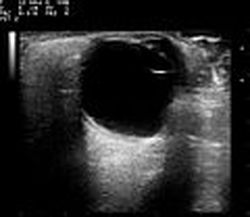
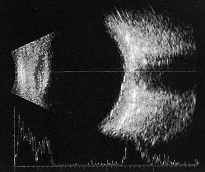
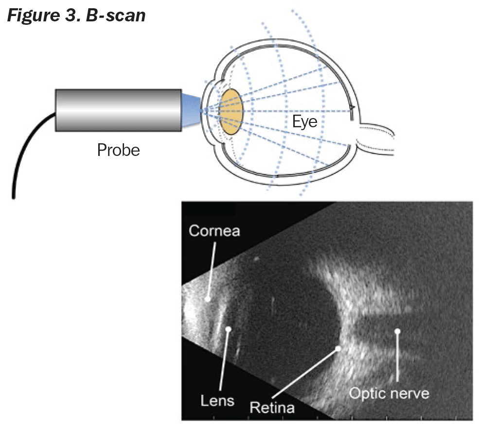
B-scan ultrasound uses high frequency soundwaves that are transmitted from a probe/transducer into the eye.

B scan. This signal is then reconstructed into a two-dimensional image on a monitor. Easy to set up defaults, smooth customer interface and automatic EMR exports to decrease examination time, increase patient throughput, and increase profit. The echoes of which are represented as a multitude of dots that together form.
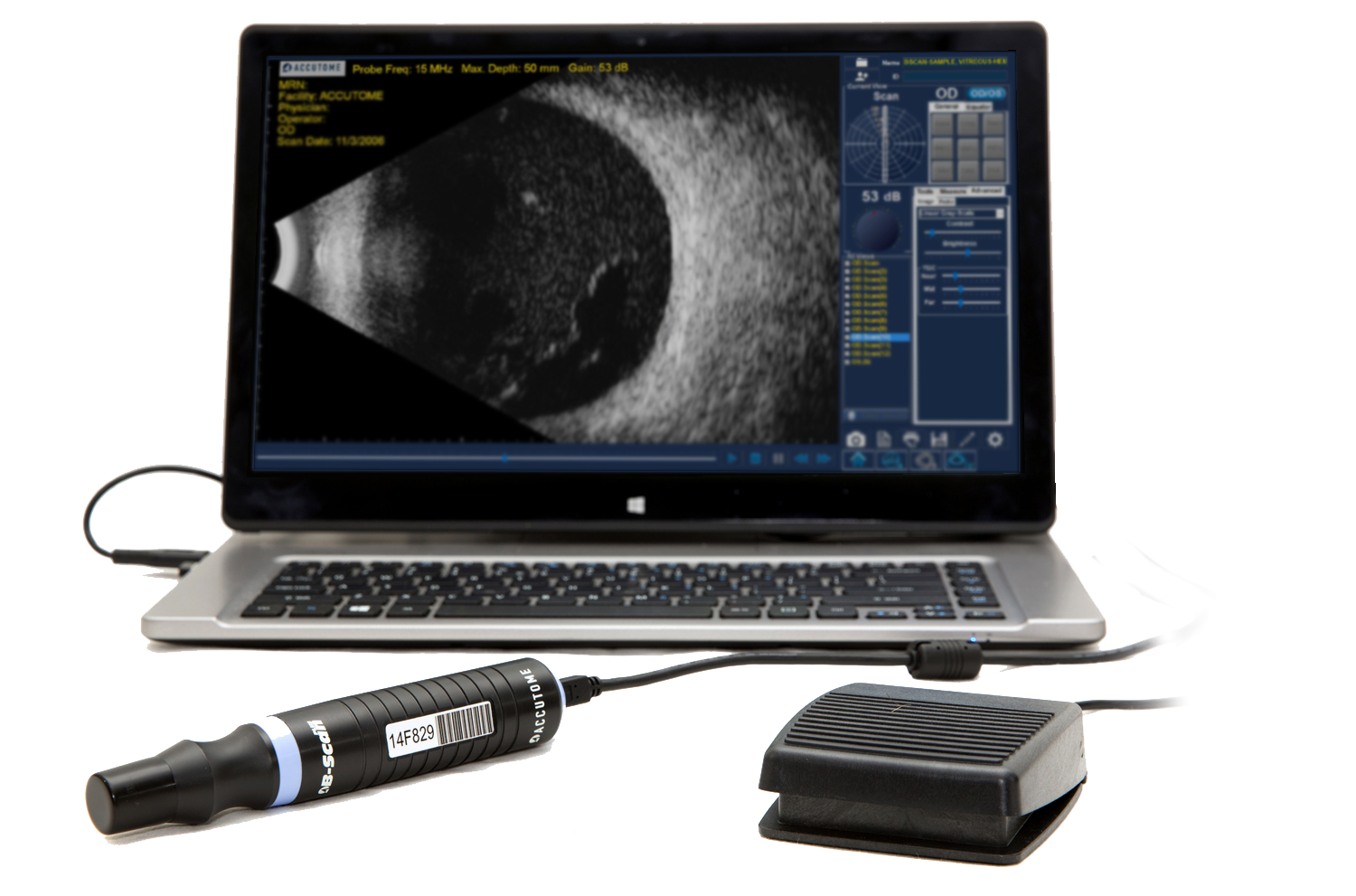
The B-scan helps your doctor see the space behind the eye. While developing the B-Scan Plus software, we visited many of the top facilities around the world. As these soundwaves strike the intraocular structures, an echo is reflected back to the probe and converted into an electrical signal.
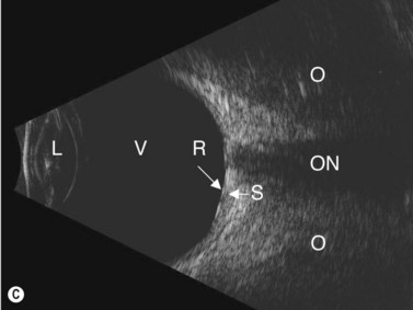
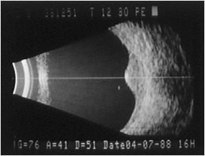
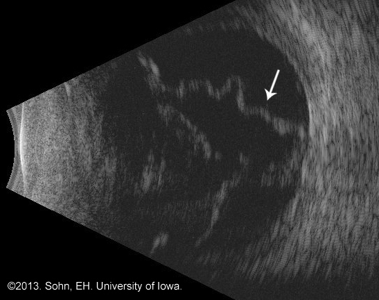
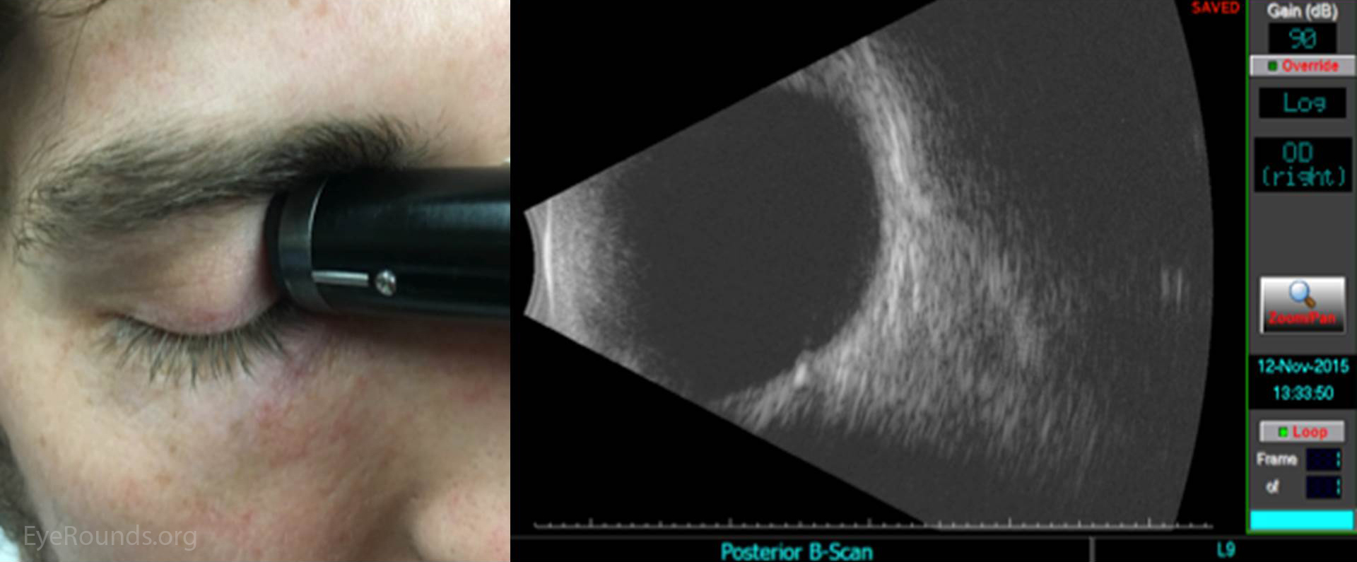
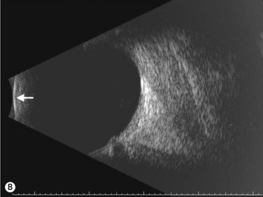
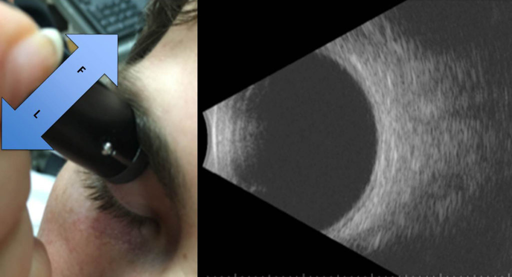
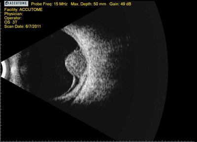
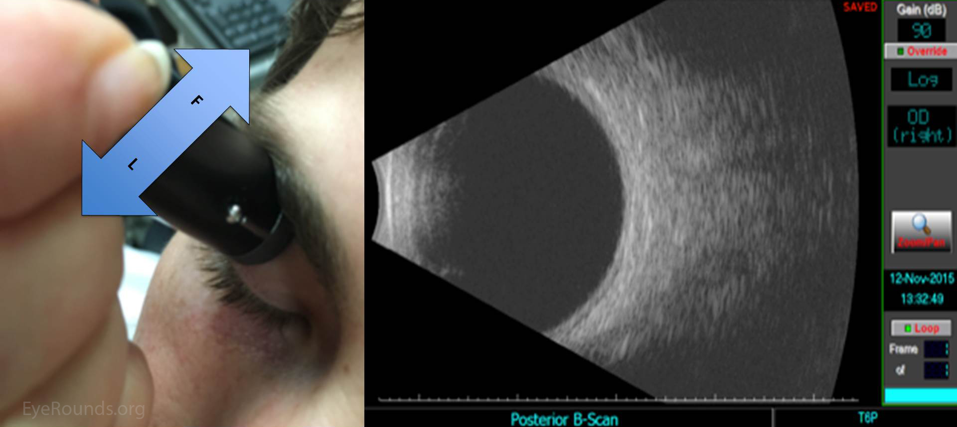
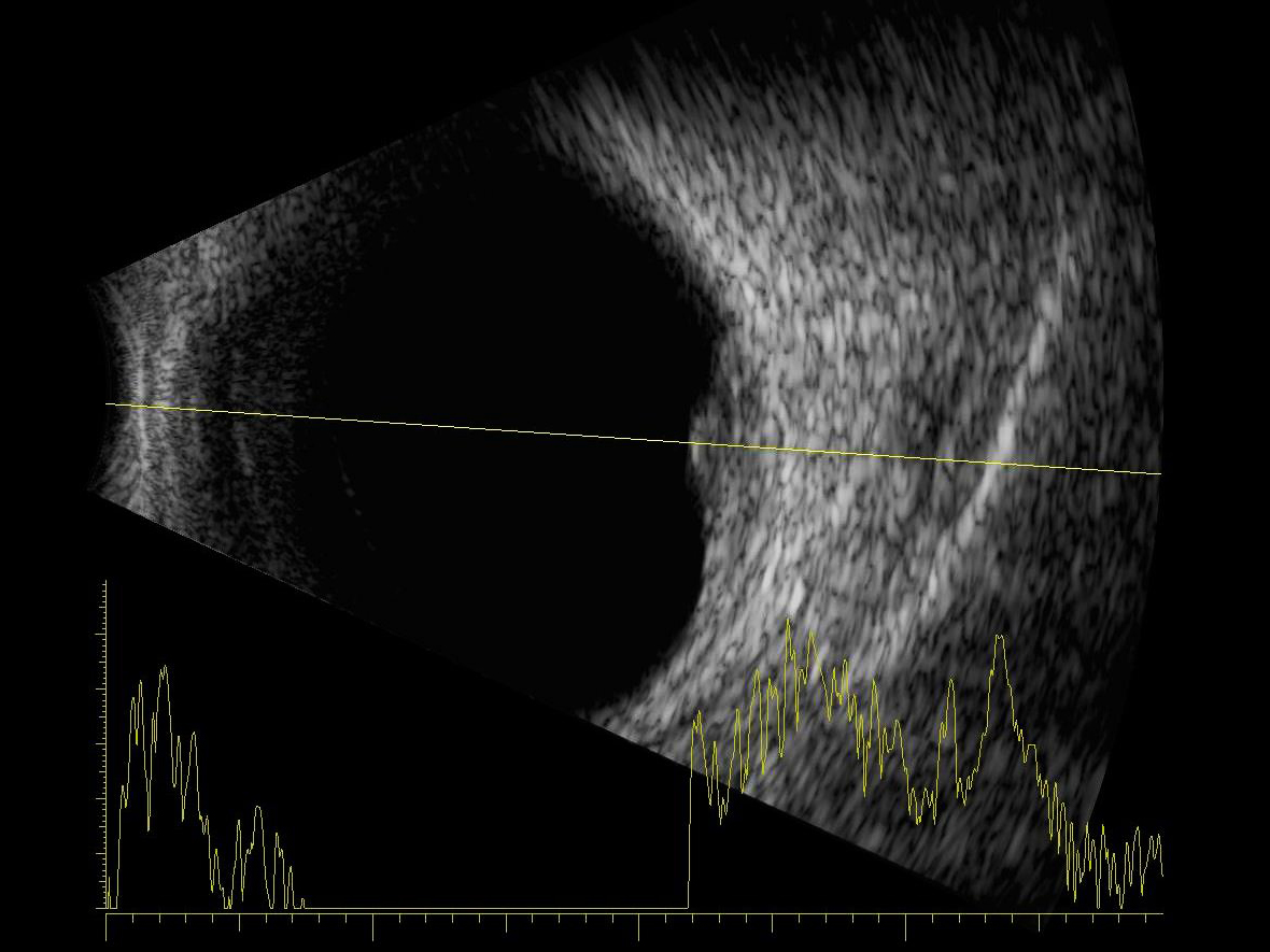
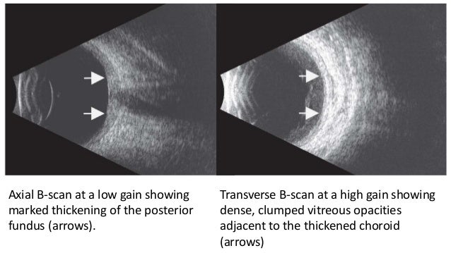
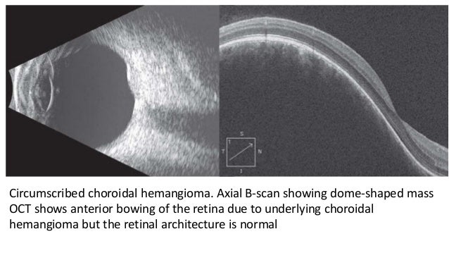
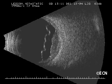
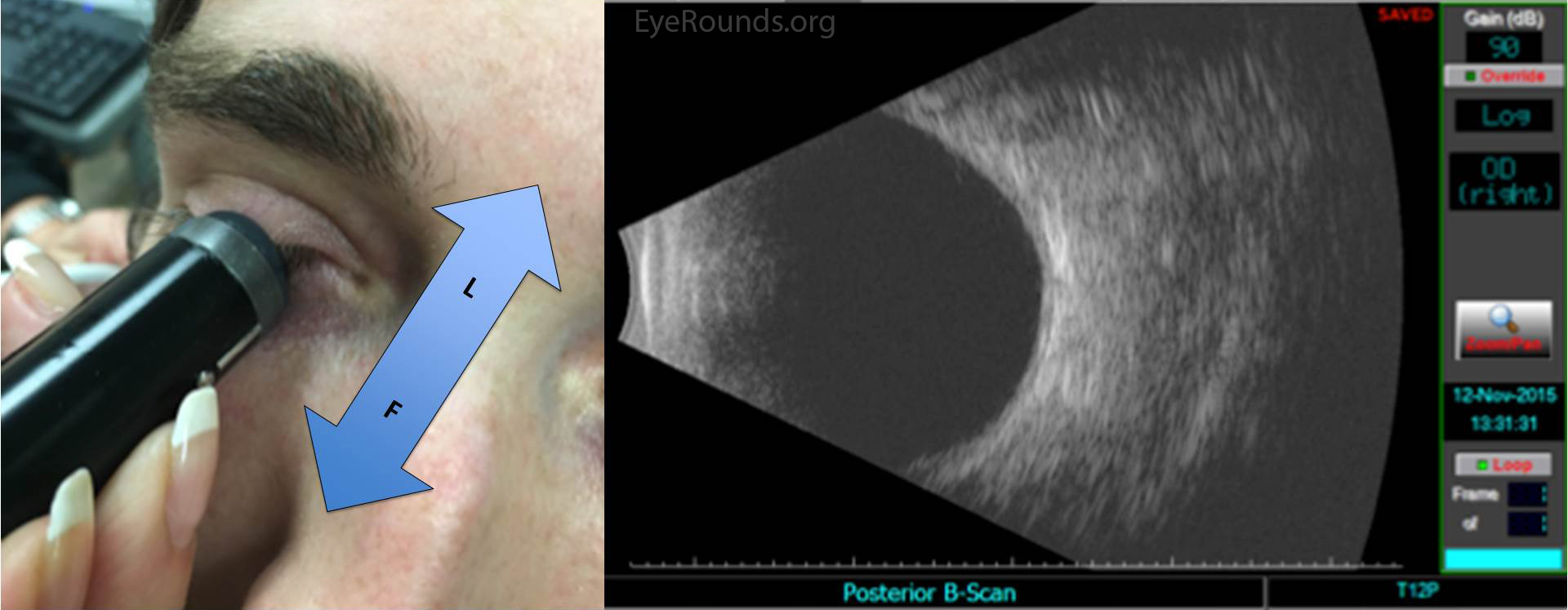
In cases of trauma or in children, B-scan can be performed over the eyelid with coupling jelly. A systematic approach should be used to acquire all images. The B-scan is useful to indicate the position of a retinal or vitreous detachment, or of an intraocular foreign body or a tumour, and for the examination of the orbit.
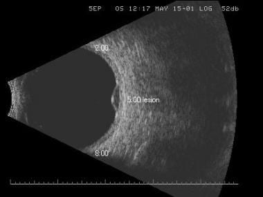
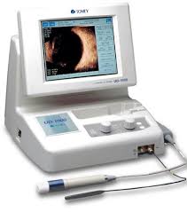
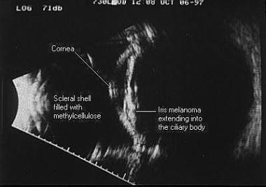
B-scan Ultrasonography, often called just B-scan or Brightness scan, offers two-dimensional cross-sectional view of the eye as well as the orbit. The results are a reliable and easy-to-use unit, which can quickly scan patients and transer information. The new Accutome Connect software allows the B-Scan Plus to integrate the option for A-scan and UBM modes for improved patient workflow.
B-scan, or brightness scan, is another method used for ocular assessment via ultrasound. The B-SCAN series helps to reduce the smuggling of drugs, weapons, mobile phones and other contraband. The B-scan is especially useful in the examination of the posterior structures of the eye when opacities prevent ophthalmoscopic examination (e.g.
A B-scan is used on the outside of the closed eyelid to view the eye. It is available in four models, to support both general and limited-use applications, in accordance with ANSI N43.17 09 guidelines. Unsurpassed compact file storage.
Cataracts and other conditions make it difficult to see the back of the eye. It can be performed directly on the anesthetized eye. In B-scan ultrasonography, an oscillating sound beam is emitted, passing through the eye and imaging a slice of tissue;.
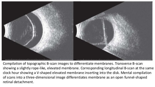
B Scan

Dealing With Hemorrhagic Choroidal Detachments Retina Today
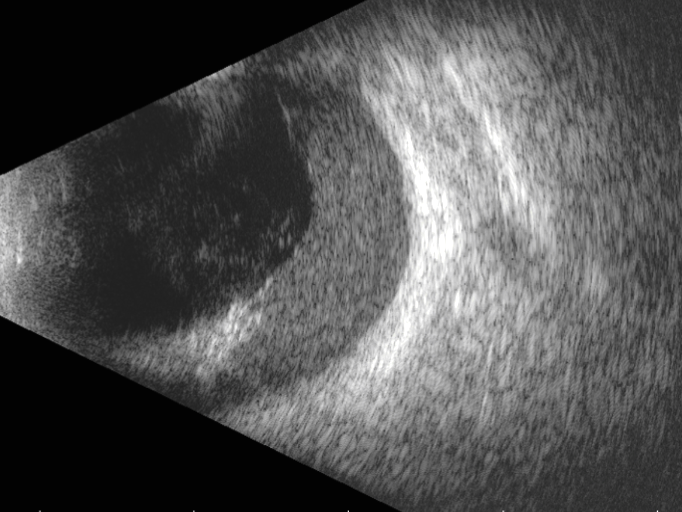
Optician
B Scan のギャラリー
Scoring An A On A B Scan

9 3 B Scan Olympus Ims
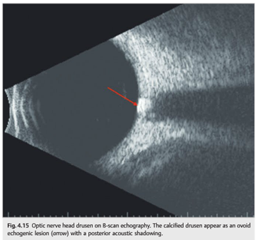
Neuro Ophthalmology Question Of The Week B Scan Ultrasound Neuro Ophthalmology

Sonomed Vupad Portable A B Scan Or B Scan
Http Www Opsweb Org Resource Collection acd 974e 40c6 A8 0dc4f627d87f Mo 1 B Affel Pdf
Q Tbn 3aand9gcqjmbcv2zymjn Lxcqvx Ch7a Qllngl Bxqehb5fu Usqp Cau

Ultrasound An Underutilized Technology In Primary Eye Care Rendia

B Scan Echography In Cases Of Confirmed Persistent Fetal Vasculature
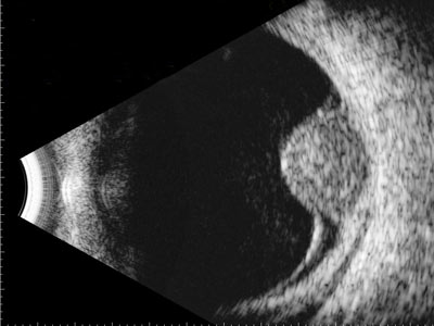
B Scan Ultrasound Retina Services Diagnostic Tests
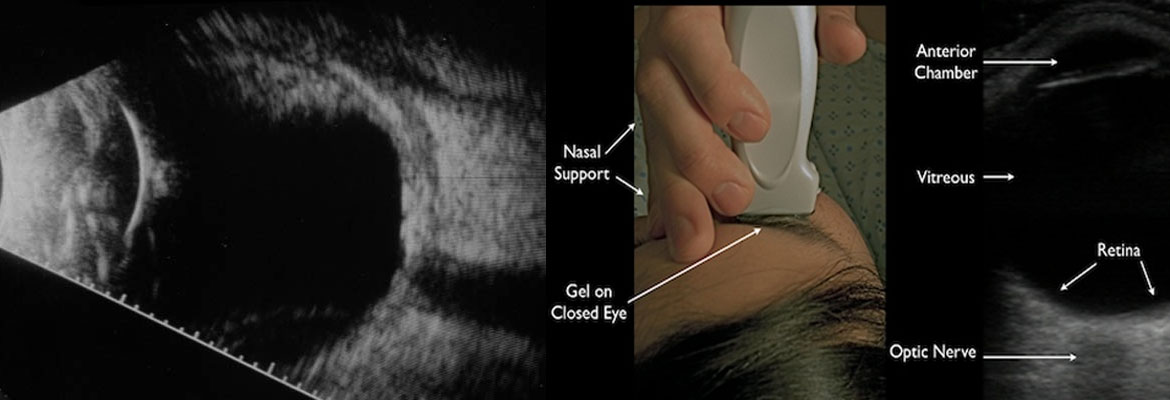
Ocular Imaging Eye Ultrasound Or B Scan Radiology Nagpur
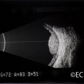
Ultrasound Images New York Eye Cancer Center
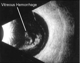
B Scan Ultrasonography Eye Day Clinic
Clinical Methods A And B Scans Radiology Key

Full Text Focused Ultrasound In Ophthalmology Opth

Ocular Ultrasound A Quick Reference Guide For The On Call Physician

B Scan Plus Ophthalmic Ultrasound Equipment
Courses And Workshops

Eyeworld Iol Preferences In Long Eyes And Their Complications

Sonomed Vupad Portable A B Scan Or B Scan
Http Www Opsweb Org Resource Collection acd 974e 40c6 A8 0dc4f627d87f Mo 1 B Affel Pdf
Retinal Detachment From One Medical Student To Another

B Scan Echography Of The Right Eye Showing Vitreous Opacities With Download Scientific Diagram
Http Www Opsweb Org Resource Collection acd 974e 40c6 A8 0dc4f627d87f Mo 1 B Affel Pdf

Predominantly Superior Retinal Tears Detected By B Scan Ultrasonography
Http Www Opsweb Org Resource Collection acd 974e 40c6 A8 0dc4f627d87f Mo 1 B Affel Pdf
Ocular Ultrasound A Quick Reference Guide For The On Call Physician
Q Tbn 3aand9gctmnri5e1q4iigy6o0syl0xs Ttzkty8gsiir O5zjzj76iyyab Usqp Cau

Optic Disc Drusen Precipitating Central Retinal Vein Occlusion In Young Bmj Case Reports
What Is B Scan Ultrasound Cairns Eye Laser Centre
Q Tbn 3aand9gctv0o6vdqftjyid3piboomigpllmeh80rpcbdjc6toapxvmwevk Usqp Cau

B Scan Ultrasonography Showing Retinal Detachment With Significant Download Scientific Diagram

Djo Digital Journal Of Ophthalmology

Choroidal Melanoma 10 Mhz B Scan The Eye Cancer Network

Ophthalmology Management The Papilledema Dilemma

B Scan Plus B Scan Biometry Ophthalmic Ultrasound Equipment
B Scan Ocular Ultrasound Overview Indications For Examination Ultrasound Principles And Physics

Mobile Usb Ophthalmic B Scan Mobile Usb Ophthalmic Device

Echography Ultrasound Eyewiki
B Scan Ultrasonography Eye Day Clinic

Use Of Ophthalmic B Scan Ultrasonography In Determining The Causes Of Low Vision In Patients With Diabetic Retinopathy Sciencedirect
B Scan Ocular Ultrasound Overview Indications For Examination Ultrasound Principles And Physics

What Should I Be Looking For When Purchasing A B Scan
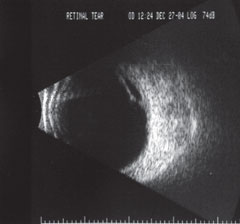
Scoring An A On A B Scan

Bemer United States

B Scan Ophthalmic Ultrasound

Role Of Ocular Ultrasound In Idiopathic Intra Cranial Hypertension

B Scan Ultrasound Images Of A Definite Retinal Tear Notes A High Gain Download Scientific Diagram
Http Www Opsweb Org Resource Collection acd 974e 40c6 A8 0dc4f627d87f Mo 1 B Affel Pdf
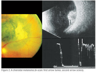
The Ongoing Role Of Ophthalmic Ultrasound

High Resolution Ophthalmic Ultrasound System For Ocular Structures Medical Design Briefs

Bright Scan Ultrasound B Scan Retina Associates Of Greater Philadelphia
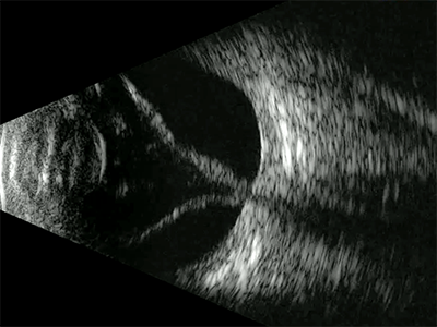
Optician
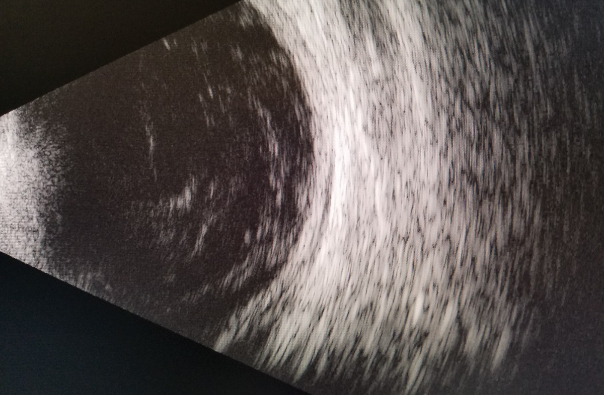
The Larry Alexander Resident Case Report Contest Cloudy With A Chance Of Retinopathy
Clinical Methods A And B Scans Radiology Key
Ocular Ultrasound B Scan Ultrasound Retina Specialists

China B Scan Ultrasound A B Scan For Ophthalmology With Trolley And Printer Md 2400s China A B Scan B Scan
Jcdr Intraocular Calcifications Imaging B Scan Ct Scan Mri Cataract Asteroid Hyalosis Retinoblastoma Meningioma

Echography Ultrasound Eyewiki

Ocular Ultrasound Retina Specialists Of Michigan

B Scan Ultrasonography Showing T Sign On The Left Eye Of The Patient Download Scientific Diagram
Ophthalmic Ultrasound Equipment B Scans Veatch Ophthalmic Instruments

Ophthalmic B Scan Ultrasound Devices Ophthalmologyweb Com

Ultrasounds Ascan B Scan Dr Michael Duplessie Kuwait Maryland

B Scan

Use Of Ophthalmic B Scan Ultrasonography In Determining The Causes Of Low Vision In Patients With Diabetic Retinopathy Sciencedirect

B Scan Ophthalmic Ultrasound Scanner अल ट र स उ ड स क नर Kiran Sonics East Godavari Id

Mobile Usb Ophthalmic B Scan Mobile Usb Ophthalmic Device

B Scan Authorstream
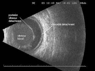
A Scan B Scan Ultrasonography Ft Lauderdale Eye Associates

Full Text Accuracy Of B Scan Ultrasonography In Acute Fundus Obscuring Vitreous Opth

Novel B Scan Technique For Distinguishing A Displaced Iol From A Retinal Mass American Academy Of Ophthalmology
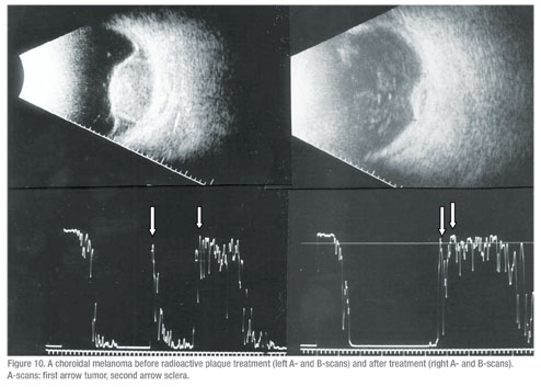
The Ongoing Role Of Ophthalmic Ultrasound
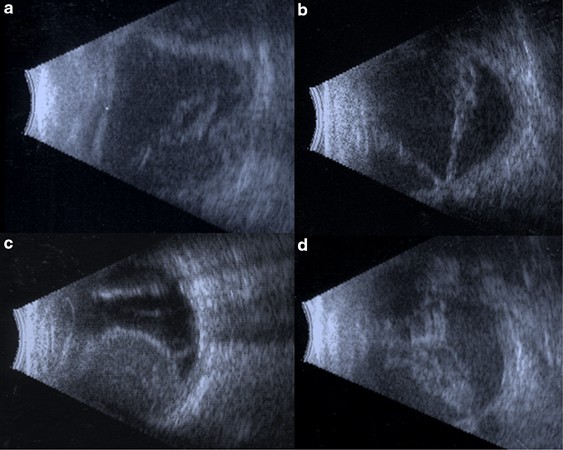
B Scan Ultrasonography Following Open Globe Repair Eye

Djo Digital Journal Of Ophthalmology

B Scan
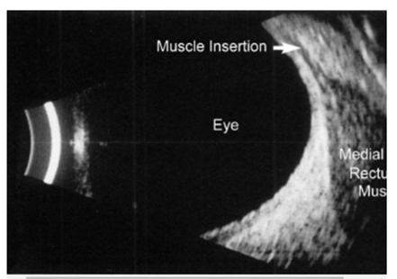
Neuro Ophthalmology Question Of The Week B Scan Ultrasound Neuro Ophthalmology

Ophthalmic B Scan Ultrasound Devices Ophthalmologyweb Com

Bright Scan Ultrasound B Scan Retina Associates Of Greater Philadelphia
B Scan Ultrasonography Troia Eye Laser Pcbeaver County Pa
B Scan Plus Ophthalmic Ultrasound Equipment
Ocular Ultrasound A Quick Reference Guide For The On Call Physician
Optician

Primary Rhegmatogenous Retinal Detachment Ento Key

Use Of Ophthalmic B Scan Ultrasonography In Determining The Causes Of Low Vision In Patients With Diabetic Retinopathy Sciencedirect
Community Eye Health Journal Caring For A And B Scans
B Scan

Bright Scan Ultrasound B Scan Retina Associates Of Greater Philadelphia
B Scan

B Scan Plus Ophthalmic Ultrasound Equipment
Q Tbn 3aand9gcrptcjxr4pvijimqs0pgjws5e2qlhetzc0ycbf252s7zdfvdpmu Usqp Cau

Moran Core Retinoblastoma

Ultrasound B Scan A Ready Reckoner For The Postgraduates

B Scan Ultrasound Of Patients Right Eye Showing The Classic Collar Download Scientific Diagram
Choroidal Metastasis 10 Mhz B Scan New York Eye Cancer Center

Compare B Scan Beye

Retinal Physician Diagnosis And Management Of Pathologic Myopia

Use Of Ophthalmic B Scan Ultrasonography In Determining The Causes Of Low Vision In Patients With Diabetic Retinopathy Sciencedirect
.jpg/image-full;max$643,0.ImageHandler)
Sturge Weber Diffuse Hemangioma And Retinal Detachment On B Scan Retina Image Bank

Ophthalmic Ultrasonography 1 Normal Anatomy Youtube
Ocular Ultrasound A Quick Reference Guide For The On Call Physician
Scoring An A On A B Scan


